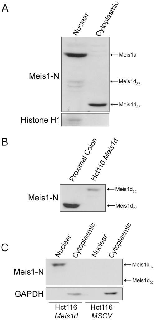
Figure 3. Posttranslational modification of Meis1d correlates with subcellular localization.
A) B6 proximal colon samples were separated into nuclear and cytoplasmic fractions and probed with the Meis1-N antibody. Histone H1 was used to confirm the subcellular fractionation. B) Western blot analysis of B6 proximal colon and Hct116 cells expressing the Meis1d ORF using the Meis1-N antibody. C) Hct116 cells were retrovirally infected with the Meis1d ORF or an empty MSCV vector. Cells were taken 72 hours post-infection, fractionated, and probed with the Meis1-N antibody. The positions of Meis1a, nuclear Meis1d (Meis1d32), and cytoplasmic Meis1d (Meis1d27) are labeled where applicable.

