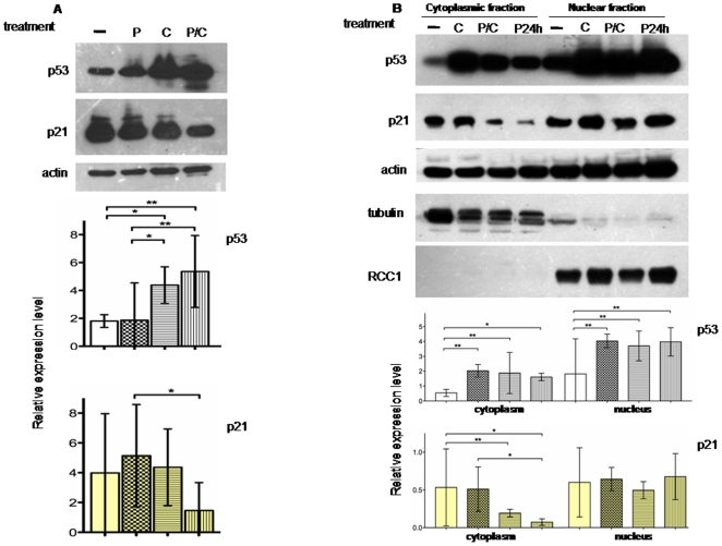
Figure 5. p21 protein is differently expressed in sub- cellular compartment.
A, Western blot analysis and relative expression level on p53 and p21 proteins after 24 hours P, C or P/C treatment in MSTO11H. The analysis reveals an increase of p53 levels after C treatment probably related to the cisplatin-induced cellular stress. Indeed p21 levels appear decreased in the P/C combined treatment. Total proteins were incubated with p21 antibody, or p53 antibody. B, Western blot analysis and relative expression level on p53 and p21 proteins in cytoplasmic and nuclear subcellular fractions. Most of the p53 protein is localized in the nucleus and there is a similar result for p21. In addition the p21 nucleus/cytoplasm ratio increases in the prolonged piroxicam pre-treatment before adding cisplatin (lanes P24h). Proteins were probed with specific cytoplasmic (tubulin) or nuclear (RCC1) antibodies to exclude fractions cross-contamination. In all the experiments, actin was used as loading control. Histograms of relative expression level refer to p53 and p21 normalized expression and derived by the analysis of three independent experiments. Statistical analysis was done as indicated in Material and Methods. -: untreated cells, P: piroxicam; C: cisplatin; P/C: piroxicam and cisplatin P24h: piroxicam and cisplatin after piroxicam pretreatment.

