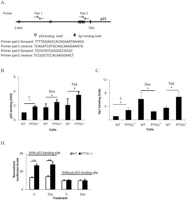
Figure 4. Sp1 and p53 regulate p21 levels and promoter activities in HCT116 WT and PTTG1−/− cells.
a) Primer pair 1 was designed to detect protein binding close to p53 motifs on p21 promoter. Primer pair 2 was used to detect protein binding close to Sp1 binding site on p21 promoter; b) WT and PTTG1−/− cells treated with control vehicle (C), doxorubicin (Dox, 0.02 uM) or TSA (0.005 uM) for 48 hours and chromatin immunoprecipitation performed using p53 antibody. Enriched chromatin was analyzed by qPCR using primer pair 1; c) WT and PTTG1−/− cells were treated with control vehicle (C), Dox (0.02 uM) or TSA (0.005 uM) for 48 hours and chromatin immunoprecipitation performed using Sp1 antibody. Enriched chromatins were analyzed by qPCR using primer pair 2; d) luciferase plasmids containing p21 promoters (with/out p53 binding motifs) were transfected into drug-treated HCT116 WT and PTTG1−/− cells. Cells were harvested and luciferase activity analyzed. Each experiment was repeated three times. Results are expressed as mean±SD, n = 3, *p<0.05, **p<0.001.

