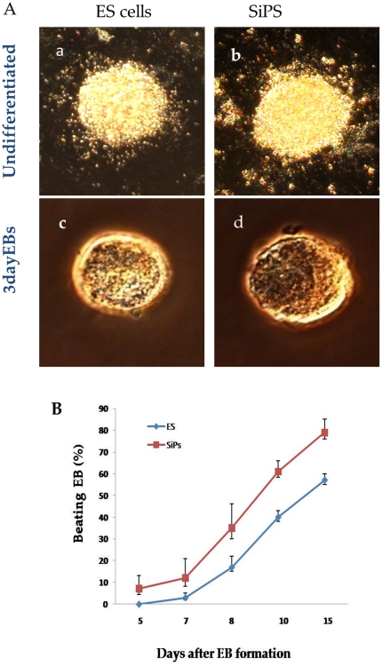
Figure 4. Embryoid bodies formation and beating pattern of murine ES cells and SiPS.
(A) Phase contrast images showing the comparable growth characteristics of undifferentiated (a) ES cells and (b) SiPS. Three days old embryoid bodies (EBs) derived from (c) ES cells and (d) SiPS. (B) Spontaneously beating EBs from the ES and SiPS over time. Data are mean ±SEM.

