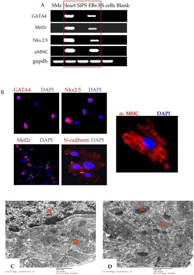
Figure 5. In vitro cardiomyogenic differentiation of SiPS.
SiPS were cultured in suspension for 3 days culture. (A) RT-PCR analysis of 5 days old spontaneously beating EBs for cardiomyocyte specific markers, Gata4, Mef2c, Nkx2.5, and α-MHC., in comparison with SMs, undifferentiated SiPS and ES cells. Mouse heart was used as positive control. Quantitative mRNA expression of Gata4, Mef2c, Nkx2.5, and α-MHC in SMs, mouse heart, SiPS, 5 days EBs and ES cells are shown in Figure S2. (B) Immunostaining of cardiac specific genes in 5 days EBs cells which were positive for cardiac specific antigens Gata4, Nkx2.5 and Mef2c (merged images with DAPI). These cells were also positive for myosin heavy chain and expressed N-cadherin (red fluorescence). (C–D) Ultra-structural features of SiPS-CPs in vitro. Transmission electron micrographs of SiPS-CPs showing typical striated sarcomeres (s) with z-lines (z), nucleus (N) and mitochondria (M) (original magnifications; A = 50000×; B = 60000×).

