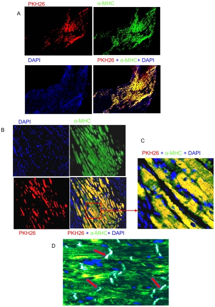
Figure 7. Histological evaluation of SiPS-CPs transplanted in the infarcted heart.
(A–D) Microscopic images from recipient mouse hearts 4 weeks post-transplantation. The surviving 5 days EBs derived cardiac progenitors are identified by PKH26 (red fluorescence). The same area was stained for myosin heavy chain (green fluorescence) and the nuclei were stained with DAPI (blue fluorescence). The superimposed photo-images identified myosin as yellow in the merged images (A–D). The transplanted PKH26 labeled cardiac progenitors (yellow fluorescence) made contact with the host myocardium (green fluorescence) through gap junctions (white), a component of intercalated disks, as indicated by red arrows. (original magnifications: A = 10×, B = 20×, C & D = 40×).

