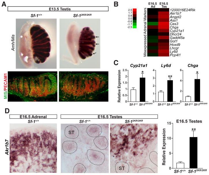
Figure 2. Misexpression of adrenal markers in Sf-12KR/2KR testes.
A. Wholemount in situ hybridization of Amh/Mis and immunofluorescent staining of MIS (green) and PECAM1 (red) in Sf-1+/+ and Sf-12KR/2KR E13.5 testes showing aberrant testes cords branching without an apparent loss of germ cells. B. Heat map showing upregulated adrenal markers in E16.5 mutant testes compared to wild type, with color scale bar shown. C. Increased expression levels of selected adrenal markers in E16.5 mutant testes. D. In situ hybridization and transcript levels (right panel) of the adrenal marker Akr1b7 in E16.5 mutant and wild type testes. ST, seminiferous tubules. See also Table S2.

