Figure 1.

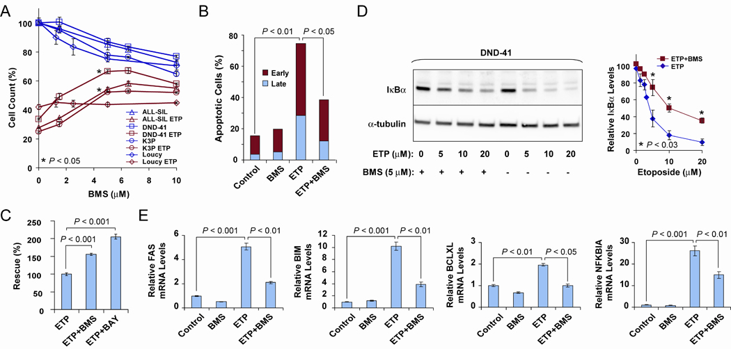
BMS counteracts etoposide effects on cell growth, IKK activation levels and NFκB target expression. A, AlamarBlue cell growth assay (25). ALL-SIL, DND-41, K3P and Loucy T-ALL cells, untreated or treated with 5 µM etoposide (ETP) and BMS at the indicated concentrations for 18 h. P values indicate significant differences in cell counts for etoposide-treated cells in the presence of 5 µM BMS (asterisks) versus etoposide alone. Measurements were performed in triplicate. Similar results were obtained for DND-41 cells (n = 5) and ALL-SIL cells (n = 2) using different times of incubation, order of etoposide and BMS addition and different sources of serum. B, Apoptosis assay (25). ALL-SIL cells were seeded at 2.5 × 10⁵/ml and early and late apoptotic cells were measured by flow cytometry after staining with annexin-PE and 7-AAD. Conditions: Control, 0.06% dimethyl sulfoxide; ETP, 10 µM etoposide; BMS, 10 µM BMS; ETP+BMS, 10 µM etoposide plus 10 µM BMS. Treatment was for 24 h. A representative graph is shown for two biological replicates. Similar results were obtained for K3P cells. C, AlamarBlue cell growth assay. DND-41 cells were treated with 5 µM etoposide and with 8 µM BMS or 4 µM BAY 11-7082 (BAY) for 18 h, as indicated. D, IKK kinase assay based on Western blot detection of IκBα degradation in DND-41 cells after 6 h treatment as indicated: ETP, etoposide. (Left) Shown is a representative blot of three independent experiments. (Right) Quantitative analysis of IκBα levels demonstrating statistically significant partial inhibition of IκBα degradation for etoposide-treated cells in the presence of 5 µM BMS (asterisks) versus etoposide alone (n = 3). E, DND-41 cells were treated as described in D and expression of NFκB targets, FAS, BIM, BCLXL and NFKBIA, was determined by qRT-PCR (19). Experiments were performed in triplicate for two biological replicates of three cell lines, ALL-SIL, K3P, and DND-41, with similar results obtained. Student’s t test for all comparisons.
