
Figure 5.
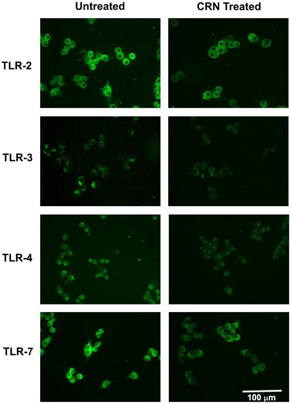
Altered expression of TLR-2, -3, -4 and -7 following exposure of RAW 264.7 cells to 0.1 mM of CRN as determined by immunofluorescent staining. Immunohistochemical staining revealing that exposure to CRN results in a decrease of TLR-2, TLR-3, TLR-4, and TLR-7 relative to the levels observed in the control-treated cells.