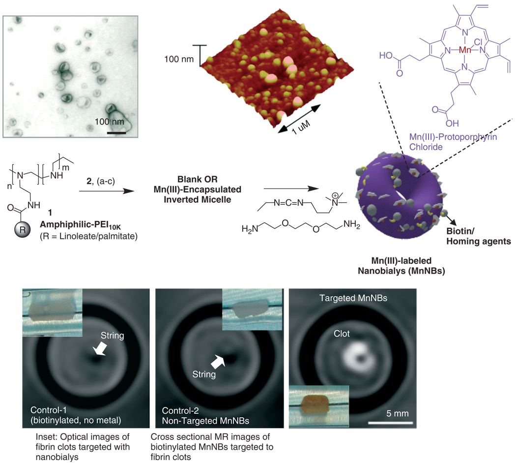
FIGURE 6.
Preparation and characterization of manganese nanobialys: (Top left) TEM (drop deposited over nickel grid, 1% uranyl acetate) and (top right) atomic force microscope (AFM) image of nanobialys (drop deposited over glass) Reaction conditions: (1) anhydrous chloroform, gentle vortexing, room temperature; (2) aqueous solution of 2 [Mn(III)-protoporphyrin], inversion, room temperature, filter using short bed of sodium sulfate and cotton; (3) Biotin-Caproyl-PE, filter mixed organic solution using cotton bed, 0.2 µM water, vortex, gently evaporation of chloroform at 45°C, 420 mbar, 0.2 µM water, sonic bath, 50°C, 1/2 h, dialysis (2 kDa MWCO cellulose membrane) against water. (Bottom) MRI images of fibrin-targeted nanobialys (right) or control nanoparticles (bottom left) and (bottom middle) bound to cylindrical plasma clots measured at 3 T.

