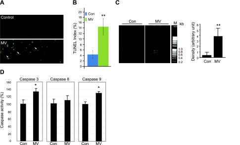
Figure 1.
MV results in DNA fragmentation and activates caspase 3 and 9 in human diaphragm. A) TUNEL staining was performed on cryosections of control and ventilated human diaphragm. Fragmented genomic DNA was labeled with FITC-conjugated dUTP; positive signals appear green. Myonuclei are visualized by DAPI staining (blue). Note that the positive TUNEL staining signals (green) are localized in nuclei stained by DAPI (blue). B) TUNEL-positive nuclei were counted and normalized to total myonuclei; ratio is shown as the TUNEL index. Control (Con), n = 9; MV, n = 10. C) DNA fragmentation was measured by PCR-based detection and visualized by electrophoresis in 2% agarose gel. Density of the PCR products was quantitated with ImageJ and normalized to the total DNA input. M, DNA markers. Control, n = 5; MV, n = 8. D) Caspase 8 and 9 enzymatic activities from control and MV human diaphragm lysate were measured by fluorometric assay. Results are presented as relative fluorescence units after normalization to total protein amount. Control, n = 7; MV, n = 9. *P < 0.05; **P < 0.01.

