Figure 3.
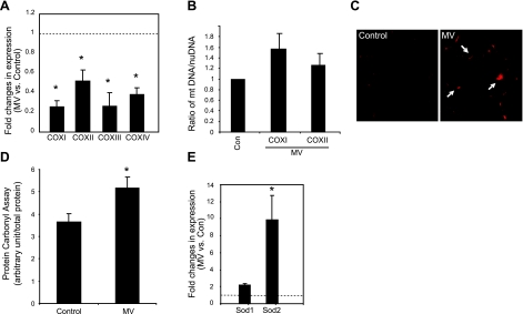
MV induces oxidative stress and impairs mitochondrial function in human diaphragm. A) Total RNA was extracted from ventilated and control human diaphragm, and quantitative real-time PCR was performed. Fold changes of COX gene expression levels were calculated as MV over control after normalization to γ-actin levels. Note that expression levels of COX I, II, III, and IV genes in ventilated human diaphragm are reduced. Control (Con), n = 7; MV, n = 9. B) DNA from ventilated and control human diaphragm was extracted, and real-time PCR was performed to examine the levels of the genomic DNA of the β-actin gene as well as the mitochondrial DNA levels of the COX I and II genes. Ratio of the COX I or II DNA levels vs. β-actin nuclear DNA levels was used to indicate relative mitochondrial content. Note that mitochondrial DNA content does not change with MV. Control, n = 5; MV, n = 5. P = 0.15. C) DHE staining was performed on cryosections of ventilated and control human diaphragm. Positive DHE staining shows as red (arrows). D) Carbonyl assay was performed on protein extracts from ventilated and control human diaphragm. Light unit of the carbonyl assay result was normalized to total protein input. Control, n = 7; MV, n = 9. E) Real-time PCR was used to quantitate the expression levels of the cytosolic and mitochondrial antioxidant genes, SOD1 and SOD2, respectively. Note that only SOD2 is significantly elevated with MV. Control, n = 7; MV, n = 9. *P < 0.05.

