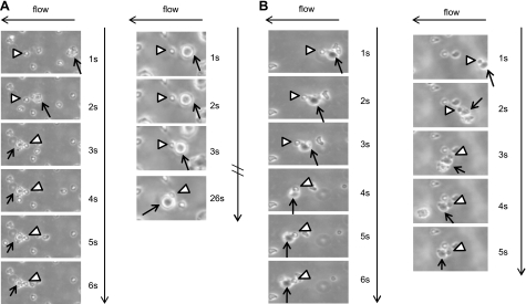
Figure 7.
Interactions between L-aptamer-MSCs and neutrophils on a P-selectin substrate under flow conditions at 0.25 dyn/cm2. Note that neutrophils and MSCs, ∼8 and ∼20–25 μm in diameter, respectively (determined by examining pure populations of neutrophils and MSCs by microscopy), can be easily distinguished from each other by size, which was confirmed by immunostaining (Supplemental Fig. 3C, D). Representative examples of an adherent neutrophil (arrowhead) capturing a flowing MSC (arrow) (A) and a neutrophil (arrowhead) complexed with MSCs (arrow) first in the flowing stream and then tethered onto the P-selectin surface (B). Note that once captured, MSCs always shift their position to the left of the immobilized neutrophils due to the flow direction (from right to left in this case), which clearly demonstrates that the capture of MSCs on P-selectin was mediated by binding of neutrophils to the P-selectin-coated substrate vs. MSCs binding to the P-selectin-coated substrate.

