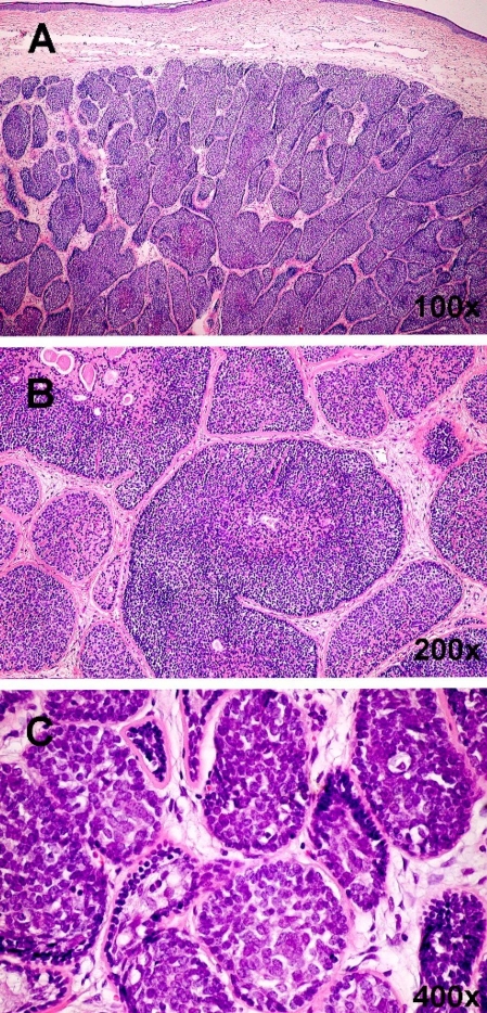
Figure 6.
Cylindroma (H&E). The pattern of cylindroma consists of dermal nests, lobules or cords of basaloid tumor cells (A). Dilated thin-wall blood vessels corresponding to telangiectasia clinically are present between the tumor mass and epidermis. Lobules are surrounded by a dense, intensely eosinophilic hyaline membrane (B). Similar eosinophilic material is present inside the nests admixed with tumor cells. The pattern formed by these almost interlocking nests has been compared to a jigsaw puzzle. Within the nests are two cell types: palisaded small cells with oval dark nuclei situated peripherally and large cells with prominent vesicular nuclei located intralobularly (C).

