
Fig. 2.
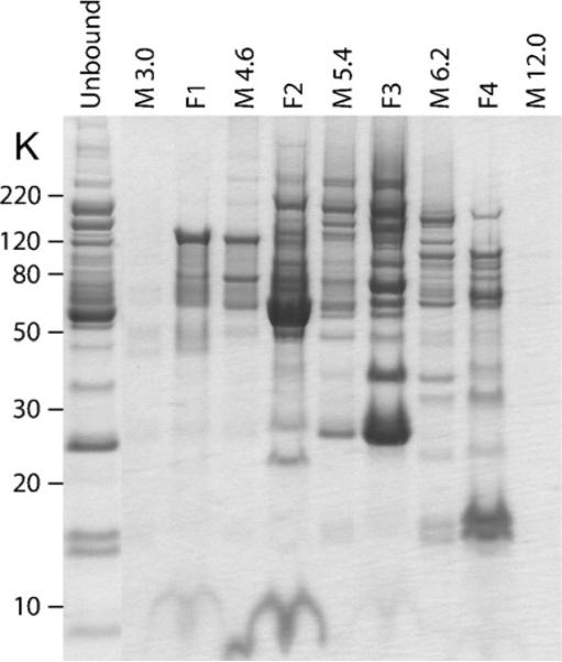
MicroSol IEF fractionation of a depleted mouse plasma sample. Depleted plasma proteins (unbound) were separated by MicroSol IEF using a four-chamber configuration. The membrane extracts (M) and the pH values are indicated. The four MicroSol IEF fractions are indicated by F1–F4. Samples were run on a 10% NuPAGE® Bis–Tris gel with MES buffer and stained with colloidal blue. A good separation is evident from the presence of unique protein bands in each of the MicroSol IEF fractions.