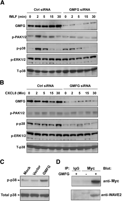
Figure 5. GMFG regulates p38 and PAK phosphorylation and interacts with WAVE2 in dHL-60 cells.
(A and B) Immunoblot analysis of the phosphorylation (p) of PAK1/2, p38, and ERK1/2 at the indicated times after fMLF (100 nM) or CXCL8 (100 ng/mL) stimulation of dHL-60 cells expressing negative-control siRNA or GMFG siRNA. Equal amounts of total cellular lysates were compared using total p38 antibody (T-p38). (C) dHL-60 cells were mock-transfected or transiently transfected with His-tagged GMFG plasmid or empty vector for 24 h and then lysed. Cell lysates were subjected to immunoblot analysis using antibodies against phosphorylated p38. Equal amounts of total cellular lysates were compared using total p38 antibody. (D) Lysates prepared from dHL-60 cells transfected with Myc-tagged GMFG (+) or empty vector (–) were immunoprecipitated (IP) with anti-Myc antibody or IgG control, and then the immunoprecipitated proteins were subjected to immunoblot analysis with anti-WAVE2 antibody. The same blot was stripped and reprobed with anti-Myc antibody. All figures are representative of at least three independent analyses.

