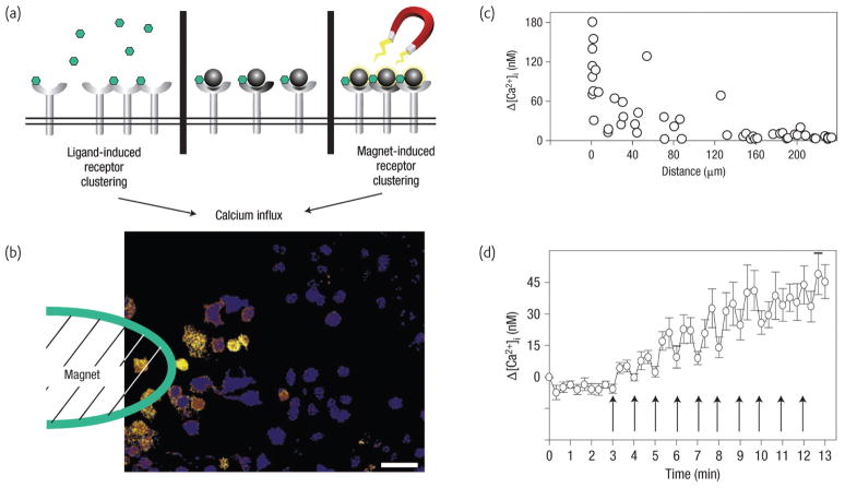
Fig. 9.
Nanomagnetic control of receptor signal transduction. a) The biochemical mechanism that stimulates downstream signalling (left) involves the binding of multi-valent ligands (represented by green hexagons) that induce oligomerization of individual IgE/FcεRI receptor complexes. In the magnetic switch, monovalent ligand-coated magnetic nanobeads (dark grey circles), similar in size to individual FcεRI receptors, bind individual IgE/FcεRI receptor complexes without inducing receptor clustering (centre). However, applying a magnetic field that magnetizes the beads and pulls them into tight clusters (right) rapidly switches on receptor oligomerization and calcium signalling. b) The pseudocoloured microfluorimetric image shows the local induction of calcium signaling (yellow) in cells near the tip of the electromagnet within 20 s of the field being applied. Scale bar is 50 μm. c) Quantification of peak changes in intracellular calcium relative to time 0 measured within individual cells during a 1-min pulse of applied magnetic force as a function of the distance of the tip from the cell surface. d) Effect of a rapid cyclical magnetic stimulation regimen (40 s on, 20 s off) on intracellular calcium signaling. (Reprinted with permission from44. ©NPG 2008).

