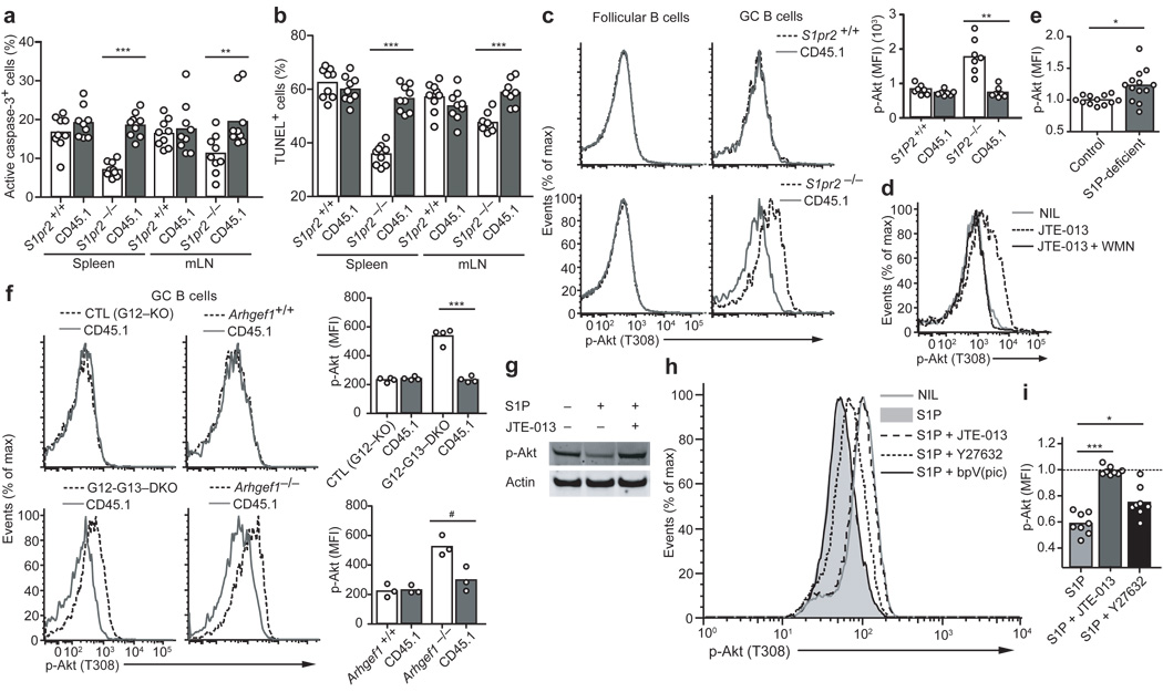
Figure 2. Apoptosis resistance and increased Akt activation in S1P2, G12–G13, and p115RhoGEF-deficient GC B cells.
Frequency of GC B cells with activated caspase-3 (a) or fragmented DNA detected by TUNEL assay (b) in chimeras reconstituted with mixtures of S1pr2+/+ or S1pr2−/− (CD45.2) and wild-type (CD45.1) BM. The mice were immunized with SRBCs and analyzed after 6–8 days. Data are from 3 experiments (n = 8–9). (c) Flow cytometric analysis of P-Akt T308 in follicular and GC B cells from mixed chimeras. Right panel shows mean fluorescence index (MFI) of P-Akt T308 in the indicated GC B cell populations (n = 7). (d) P-Akt T308 analysis in wild-type GC B cells from spleen suspensions treated with JTE-013 or JTE-013 and wortmannin (WMN) for 30 min immediately ex vivo (n = 3). (e) Graph showing P-Akt T308 MFI of mLN GC B cells from Mx1-cre+Sphk1f/− or f/fSphk2−/− (S1P-deficient) mice, relative to the average of the controls (n = 12–13, pooled from 6 experiments). (f) P-Akt T308 analysis in GC B cells deficient in p115RhoGEF or both G12 and G13, compared to littermate control cells and wild-type (CD45.1) cells, from mixed BM chimeras. The G12–G13 DKO littermate control was G12-single deficient. Right panels show a summary of P-Akt T308 MFIs in GC B cells from mixed chimeras (data are representative of 2 experiments). (g) Western blot for P-Akt S473 in Ramos cells treated for 5 min with S1P in the presence or absence of JTE-013 (representative of 3 experiments). (h) P-Akt T308 analysis in Ramos cells treated for 5 min with S1P (10nm) alone or in the presence of the indicated inhibitors. Y27632, 10µm; JTE-013, 10µm; bpV(pic), 500nm. (i) Relative P-Akt T308 MFIs in Ramos cells treated with the indicated conditions, compared to untreated (dashed line) (n = 8, pooled from 8 experiments). # P ≤ .05, * P ≤ .01, ** P ≤ .001, *** P ≤ .0001.

