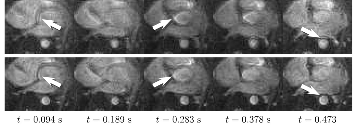
FIG. 7.
Sequential axial slices from a real-time cardiac study corrected using the image convolution method. The deblurring is noticeable in the aorta and atrial walls with some residual blurring partially due to motion. The 5 mm slices were obtained at X = (17.2,−0.8,−50.0) mm. The top row is the original image series, and the bottom row is the corrected image series using both the field map and the concomitant gradient fields.

