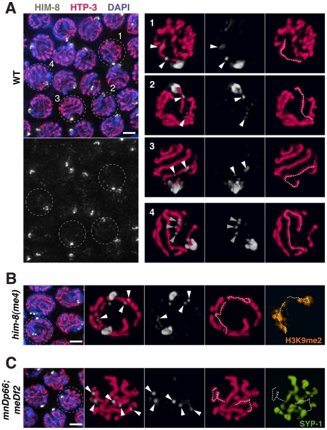
Figure 10. HIM-8 localizes to chromosome sites outside the X Pairing Center.
(A) Mid-pachytene nuclei in wild type show one major focus of HIM-8 (white) staining corresponding to the paired X PCs, as well additional fainter HIM-8 speckles elsewhere in the nucleus. Images on the left are projections of deconvolved optical sections spanning the full nuclear depth. Scale bar, 2 µm. Images on the right are 3-D surface renderings comprising half the depths of nuclei 1–4. These images show HIM-8 speckles localizing to discrete sites along the lengths of the X chromosome and autosomes, with paths of the chromosomes visualized by immunostaining of chromosome axis marker HTP-3 (pink). While there is generally at least one speckle adjacent to each chromosome axis, the most prominent HIM-8 speckles are found along the axis of the X chromosome and at one end of an autosome. Nuclei 1–3 show the typical localization of two prominent HIM-8 speckles (white arrowheads) along the axis of the X chromosome (white dotted trace); 3 or more prominent speckles are sometimes detected on the X. Nucleus 4 shows a cluster of HIM-8 speckles (grey triangles) characteristic of one end of an autosomal axis (grey dotted trace). (B) Analysis of HIM-8 localization in the him-8(me4) mutant. Mid-pachytene nuclei show two major foci of HIM-8 marking the unpaired X PCs; in this field, the HIM-8 foci are well separated in the Z dimension in all nuclei. Scale bar, 2 µm. The indicated nucleus is depicted in 3-D as in (A). HIM-8 speckles localize adjacent to the axes of the unpaired X chromosomes, which are highlighted by immunostaining of H3K9me2 (orange), which concentrates on unsynapsed X chromosomes [48]. (C) Analysis of HIM-8 localization in the meDf2 homozygote. Mid-pachytene nuclei show one major focus of HIM-8 staining corresponding to the paired mnDp66 chromosomes, which contain a copy of the X-PC region fused to chromosome I. The meDf2 X chromosomes, which lack the X-PC and thus do not have an associated major HIM-8 focus, are unpaired. Scale bar, 2 µm. The indicated nucleus is depicted in 3-D as in (A). (Note: the major HIM-8 focus on the mnDp66 chromosome is in the half of the nucleus not depicted in the 3-D rendering.) The axes of the unpaired X chromosomes are identified by lack of associated immunostaining for SC central region protein SYP-1 (green). Although these X chromosomes lack PCs and the associated major HIM-8 foci, HIM-8 speckles still localize along the unpaired X chromosome axes. In addition, the paths of the X chromosome axes in this nucleus are coiled (left) or folded (right), although they appear comparable in length to those seen in the him-8(me4) mutant. This type of organization is common for X chromosomes in meDf2 homozygotes and may account for the fact that meDf2 X chromosomes have relatively compact ovoid territories despite exhibiting increases in the number of discernable painted chromosome segments.

