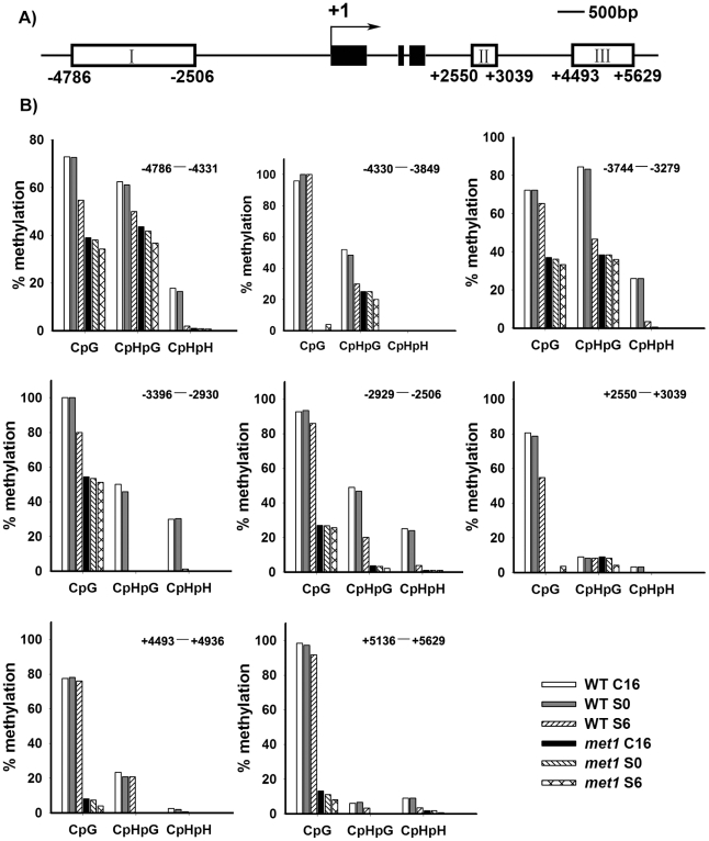
Figure 4. Analysis of WUS methylation through bisulfite genomic sequencing.
A) A diagram of WUS structure, with +1 as the transcription start site and rectangles representing the methylated region I, II and III. B) Cytosine methylation at region I, II and III of WUS was determined by bisulfite genomic sequencing. Genomic DNA methylation status of WUS is shown in calli of the wild type on CIM for 16 days (WT, C16) and for 20 days (WT, S0), and on SIM for 6 days (WT, S6). Calli of met1 are incubated on CIM for 16 days (met1, C16) and for 20 days (met1, S0), and on SIM for 6 days (met1, S6). H represents A, T or C.

