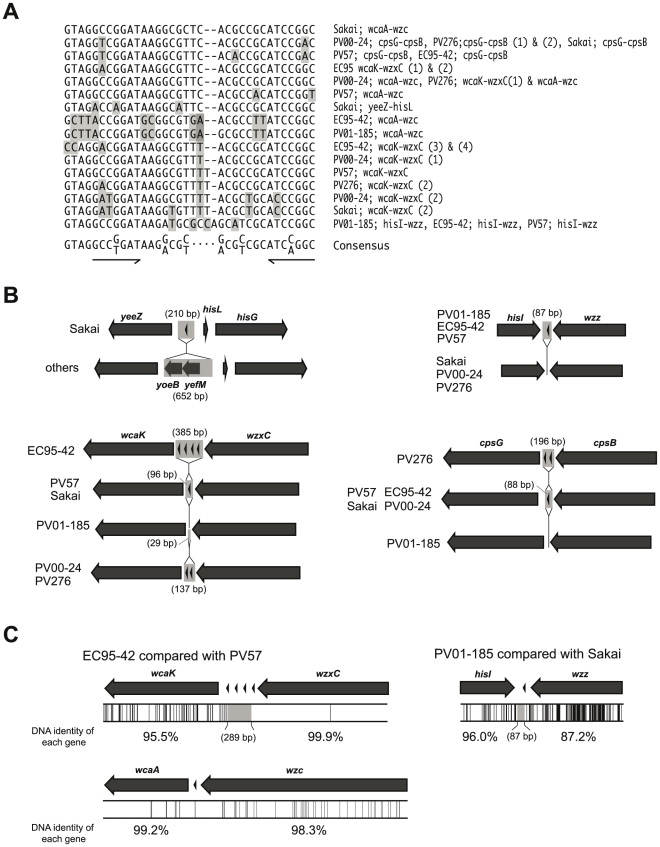
Figure 4. Schematic drawing of REP sequence-containing regions of O157-antigen biosynthesis gene cluster flanking regions.
(A) Sequence alignment of the REP sequences located in the O157-antigen gene cluster flanking regions. The consensus sequence is derived from previously published data [40]. The palindromic motif is underlined. The non-consensus sequences were highlighted. (B) Four regions showing insertion and/or deletion of fragments including REP sequence(s); yeeZ-hisG, hisI-wzz wcaK-wzxC and cpsG-cpsB are compared between strains. REP sequences are indicated by arrowheads and gray boxes indicate missing regions on each of the compared strains. (C) The nucleotide sequences from wcaK to wzxC and from wcaA to wzc on EC95-42 are compared with those of PV57, and the sequences from hisI to wzz on PV01-185 are compared with those of Sakai. Locations of SNPs by pairwise sequence comparison are indicated by vertical lines (lower panel).

