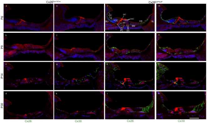
Figure 3. Time course of connexin immunoreactivity in the sensory epithelium of Cx26Sox10Cre mice.
Maximal projection rendering of two consecutive midmodiolar confocal optical sections taken at 1 µm intervals in the basal cochlear turn of Cx26Sox10Cre mice (panels a–h) and control Cx26loxP/loxP mice (panels i–p) at P6, P9, P14 and P30. Expression of Cx26 (panels a–d, i–l) and Cx30 (panels e–h, m–p) was detected with selective antibodies (green) nuclei were stained with DAPI (blue) and actin filaments with Texas red conjugated phalloidin (red). TCC, tall columnar cells forming a transient structure, also known as Kölliker's organ; CC, cuboidal cells that replace TCC during the first two postnatal weeks; DC, Deiters' cells (also known as outer phalangeal cells); IHC, inner hair cell; IS, inner sulcus; SLi, spiral limbus; OHC, outer hair cells; OS, outer sulcus; PC, pillar cells forming the tunnel of Corti (TC); NS, Nuel's space; V, vas spiralis; BM, basilar membrane; SL, spiral ligament; scale bar, 50 µm.

