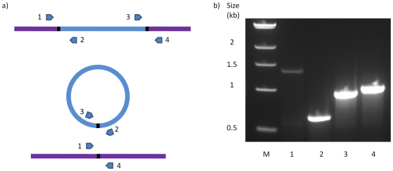
Figure 1. Detection of excision of the elements from the genome.
a) Schematic of primer binding sites on the element and the genome. The chromosomal region is shown in lilac and the element in blue, the circular form of the element is also shown (centre), as is the regenerated target after excision (bottom). Oligonucleotide primers and their direction of priming are represented by arrows. Primer pair 1+4 will detect the empty target site, primer pair 2+3 will detect the circular form of the element, primer pairs 1+2 and 3+4 for detection of the junctions between the genome and the element. b) PCR products run on 1% agarose gel for CTn5 excision from 630 genomic DNA. Lane 1; primers 2+3, lane 2; primers 1+4, lane 3; primers 3+4, lane 4; primers 1+2.

