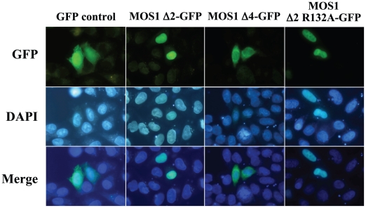
Figure 4. Comparisons of fluorescence patterns between MOS1 Δ2-GFP and two variants.
The GFP fluorescence patterns are analyzed in HeLa cells transfected with plasmids expressing only GFP or MOS1 Δ2-GFP as controls, MOS1 Δ4-GFP or MOS1 Δ2 R132A-GFP variants. The top panels show GFP fluorescence, the middle panels show the nuclear genomic DNA staining by DAPI, the bottom panels correspond to merge pictures. The observation of aggregates was already reported in the literature with the transposon protein MURB (about 26 kDa; [20]). This accessory protein is encoded by the plant transposon MuDR and assembles aggregates in the cytoplasm when it has an important expression rate.

