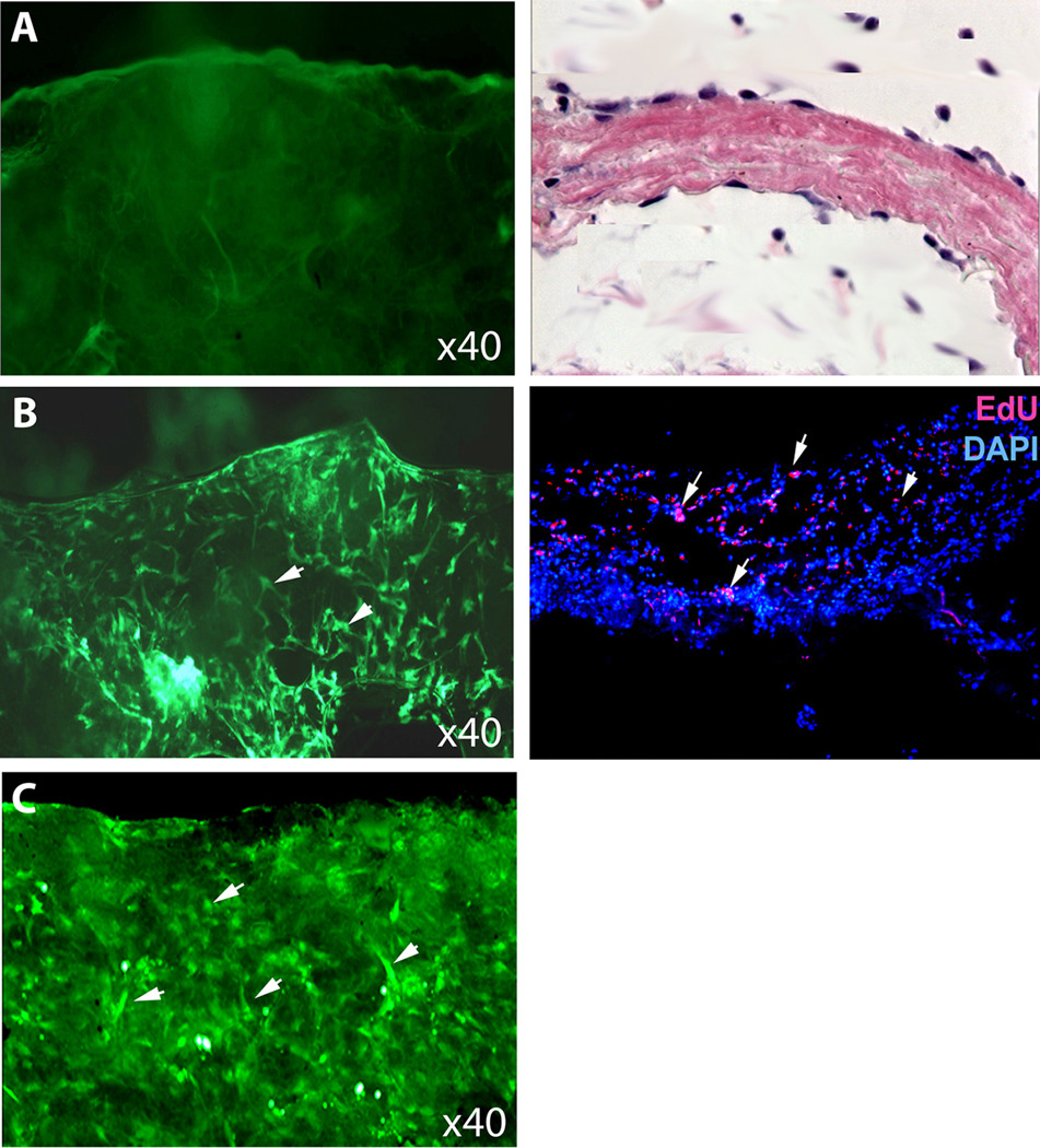
Fig. 2.
Seeding with ADSC. A. The decellularized matrix was devoid of cells as indicated by calcein stain (green). B. Twenty-four h after seeding 55% of the surface was covered with ADSCs (arrowheads). C. One week after seeding 90% of the surface was covered with ADSCs (arrowheads). D. The seeded matrix was transplanted into subcutaneous space, which was examined by HE staining 10 days later. E. Another transplanted matrix was examined by EdU staining for the presence of seeded ADSCs (arrowheads) and by DAPI staining for cell nuclei.

