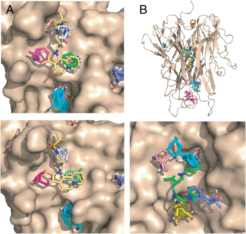
Fig. 3.
Mapping results for Zip and TNF-α. A. Mapping of ZipA. (Top) Unliganded ZipA (PDB ID code 1f46). CS1 (cyan, 17) is not in the ZipA/FtsZ peptide interface and does not overlap with the inhibitor. The consensus sites in the interface are CS8 (green, 6), CS9 (gray, 5), and CS11 (magenta, 3). (Lower) ZipA cocrystallized with compound 5 (Fig. S1). (PDB ID code 1s1s). The consensus sites in the interface are CS4 (gray, 10), CS8 (green, 4), and CS10 (magenta, 2). B. Mapping of TNFα. Top panel: Intact TNFα trimer. The only consensus site in the TNFα-TNFR1 interface is CS7 (orange, 5). The hot spots CS1 (cyan, 22), CS2 (magenta, 20), CS3 (19, yellow), CS4 (salmon, 7), and CS6 (blue, 6) are in the interior of the protein. (Lower) Results for the A and B chains of TNFα obtained by removing chain C from the trimeric structure (PDB ID code 1tnf). CS2 (cyan, 19) overlaps with the trifluoromethylphenyl indole moiety of compound 6 (shown in green), CS3 (yellow, 19) is close to K98 and Y119, CS4 (salmon, 10) is near Y59 and Y151, and CS6 (blue, 9) overlaps with the dimethyl chromone group.

