Fig. 3.
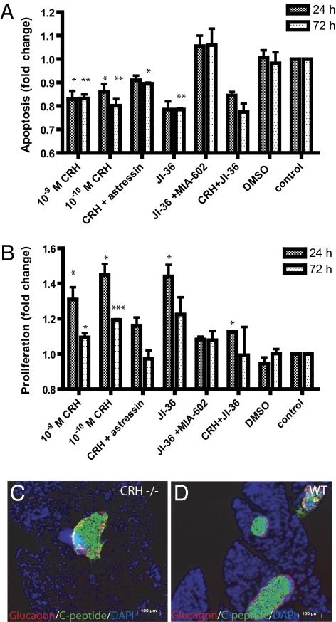
Effects of hypothalamic peptides on apoptosis and proliferation of insulinoma cells. (A) Apoptosis as indicated by activity of caspases 3/7 was significantly reduced by 19.8 ± 6.9% after 72-h cultivation with 10−10 M CRH in contrast to untreated cells. Cultivation of INS-1 cells with CRH and the CRH-antagonist, astressin, partly blocked the effect of CRH alone. The GHRH-agonist JI-36 was used as positive control, decreasing the apoptosis by 21.5 ± 4.9%. This effect could be blocked completely by the GHRH-antagonist MIA-602. The combination of CRH with JI-36 revealed 22.5 ± 4.9% reduced apoptosis (n = 3). (B) Evaluation of cell proliferation was performed by BrdU incorporation, whereby CRH (10−10 M) revealed significant increase in proliferation by 44.8 ± 10.7% after 24-h cultivation in contrast to control. Astressin revealed a blockage of the proliferative effect of CRH. The positive control, JI-36, revealed an increased proliferation of 44.0 ± 11.5%. However, this proliferative effect was blocked by the GHRH-antagonist MIA-602. Cultivation of INS-1 cells with CRH plus JI-36 showed no effect on proliferation (n = 3). CRH treatment increased dose-dependently cell proliferation and conversely decreased the rate of apoptosis in INS-1 cells, with the maximum effect at 10−10 M. DMSO was used as solvent control in same dilution as the substance. (*P < 0.05; **P < 0.01; ***P < 0.001) Immunohistochemical staining of pancreatic islets for glucagon and C-peptide indicates lower islet number in (C) CRH −/− mice than in (D) control animals (WT).

