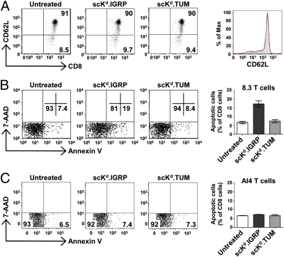
Fig. 3.
Induction of apoptosis in naive CD8+ T cells by scKd.IGRP tetramers. (A) Splenocytes from 6- to 8-wk-old 8.3 TCR-transgenic NOD mice were incubated at 37 °C for 3 h with 25 nM scKd.IGRP tetramers (red histogram) or 25 nM scKd.TUM tetramers (blue) or left untreated (filled gray), and then stained with anti-CD8 and anti-CD62L and analyzed by flow cytometry. Samples were gated on CD8+ cells. (B) As in A, except that treated and untreated splenocytes were stained with anti-CD8, Annexin V, and 7-AAD and analyzed by flow cytometry. Samples were gated on CD8+ 7-AAD− cells. (C) As in B, except that splenocytes from 6- to 8-wk-old AI4 TCR-transgenic NOD mice were used. In A–C, numbers denote the percentage of cells present in the indicated quadrants of the dot plots.

