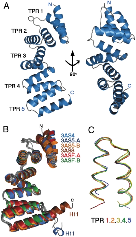
Fig. 3.
Overall MamAΔ41 structures and TPR representations. (A) The representative MamAΔ41 structure (3AS4 in cartoon drawing) contains five sequential TPR motifs. The molecule is shown in two views, related by a 90° rotation. (B) Overlay of all MamAΔ41 monomers (in cartoon drawing) reveals the high degree of structural similarity, apart from the His-tag linker sequence remaining after thrombin proteolysis (H11: ELALVPR), which appears in the 3AS5 chain A and 3AS8 structures. (C) Overlay of all five TPR units within the 3AS5 chain A structure (in ribbon drawing) shows a conserved, typical TPR fold. All structural images were prepared using PyMOL.

