Fig. 3.
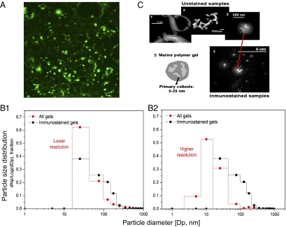
Microgels in clouds. (A) Immunostained gels. Nanometer (nanogels)- and micrometer (microgels)-sized polymer gels immunostained with a specific antibody developed toward polymeric material collected in SML and SSW. (B) Gel size distribution from two independently analyzed subsamples. B1, Relative frequency of particle number (Np) [dNp/log(dDp): delta of Np/log delta particle diameter (Dp)] in a specific size range for unstained nanogels observed with FESEM (circles) and immunostained gels observed with confocal microscopy (squares), with instruments held at a similar, lower resolution. B2, The higher resolution of the FESEM allows the measurement of nanogels smaller than 10 nm. (C) Structure of nano and microgels. (C1) Low water-soluble organic particles in the SML present large quantities of colloidal-sized nanogels (<1 μm), which annealed into microgels bigger than 3 μm. (C2) Colloidal-sized nanogels tend to present fractal structures in sizes generally <200 nm and always smaller than 1 μm in both SML and cloud samples. (C3 and C4) The gels in the cloud samples present average sizes between 200 and 700 nm, with nanogels partitioned inside of both nonstained and immunostained particles. (C5) The schematic illustration shows that colloidal-sized nanogels tend to present a fractal structure, with a diffuse organic surface.

