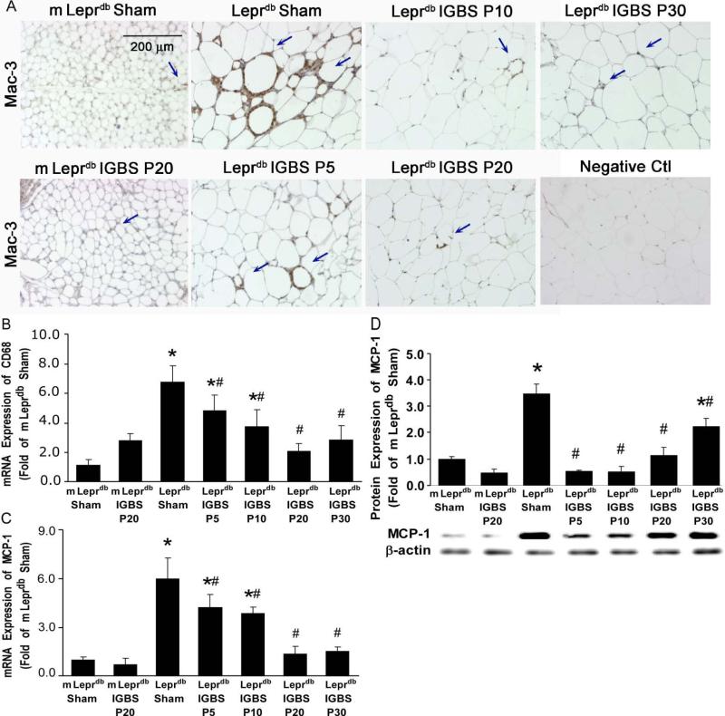
Figure 2.
IGBS reduced macrophage infiltration and MCP-1 expression in MAT of diabetic mice. A, Immunohistochemical staining shows that Mac-3 positive macrophage infiltration in MAT was higher in Leprdb+Sham versus IGBS. Data shown are representative of 4 separate experiments. B, mRNA expression of CD68 was increased in MAT of Leprdb+Sham. IGBS significantly reduced CD68 mRNA levels in MAT. The mRNA (C) and protein (D) expression of MCP-1 were increased in MAT of Leprdb+Sham, and were reduced by IGBS. Data represent mean±SEM, n=4-12 mice. *P<0.05 compared with m Leprdb+Sham surgery; # P<0.05 compared with Leprdb+Sham surgery.

