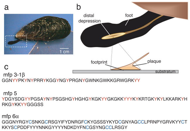
Figure 1. Byssal adhesion in the California mussel Mytilus californianus.
(a) An adult mussel attached to a mica sheet by a byssus containing 3 threads. Extended foot is making a new plaque and thread; (b) Schematic zoom of a foot viewed from the underside showing the distal depression lifting off from a completed plaque. The footprint denotes the distalmost part of each plaque in intimate contact with the surface; (c) Primary sequence of selected footprint protein variants of mfp-3 (>25 known variants), mfp-5 (2 known variants), and mfp-6 (5 known variants); the high proportion of dopa (red Y) and cysteine (blue C) in mefp-3, mfp-5 and mfp-6, respectively, is as shown.

