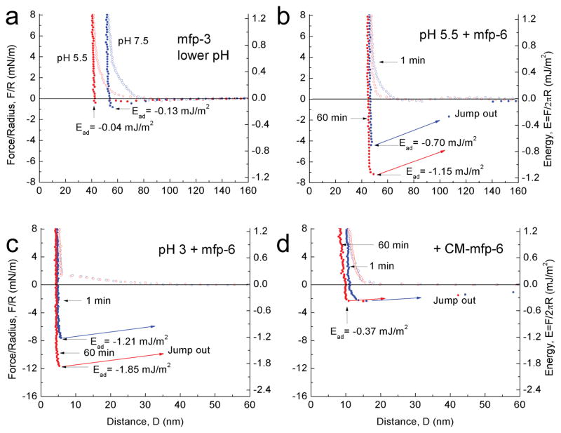
Figure 2. Adhesion of mfp-3 at different pH before and after mfp-6 addition.
In each experiment, the adhesion of symmetric mfp-3 films was determined at pH 3 before raising the pH to 7.5. (a) Simply decreasing the pH from 7.5 to 5.5 did not recover significant adhesion (see Supplementary Fig. 3) (b) After injecting mfp-6 into the gap solution at pH 5.5 (following a), an increased adhesion energy was measured after keeping the two surfaces in contact for 1 min (blue); the energy increased even more after a 60 min contact time (red). (c) As in (a), dropping the pH from 7.5 to 3 did not recover mfp-3 adhesion (not shown), however, injecting mfp-6 at pH 3 increased the adhesion energy to its highest levels at 1 min (blue) and 60 min (red); (d) Injecting S-carboxymethylated mfp-6 at pH 3 failed to recover the lost adhesion of mfp-3 cycled from pH 3 to 7.5, and back to pH 3. The experiments of (a) and (b) were repetitive, whereas (c) and (d) were de novo.

