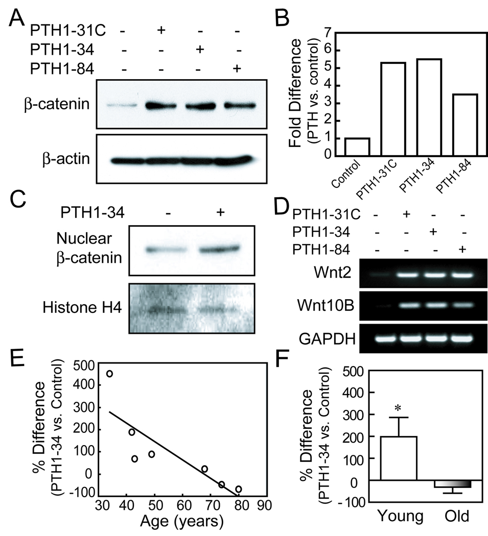
FIG. 3.
Effects of age on stabilization of β-catenin by PTH analogs in hMSCs. (A) Western immunoblot shows that after 6 hours treatment, PTH1-31C, 1–34, and 1–84 (10 nM) increased β-catenin levels in hMSCs (representative immunoblot with hMSCs from a 34-year-old female subject). (B) Quantitative data from Western immunoblots are presented as fold-difference of β-catenin with PTH treatment compared with vehicle control after normalization to each β-actin internal control. (C) PTH1–34 (10 nM) increased nuclear β-catenin in hMSCs as shown by Western immunoblot with equal loading of 10 µg in each sample. Histone H4 served as an internal control. (D) Electrophoretogram of RT-PCR products shows upregulation of Wnt2 and Wnt10B genes in hMSCs (34F), after 3 days treatment with PTH analogs (10 nM) in osteogenic medium. GAPDH was used as an internal control. (E) The effect of PTH1–34 on β-catenin in hMSCs was inversely correlated with subject age (Pearson r=−0.84, p=0.018, n=7). (F) The mean effect of PTH1–34 on β-catenin was significantly higher in the group younger than 50 years (n=4) than in the group of subjects older than 55 years (n=3) (*p<0.05, Mann-Whitney test).

