
Figure 1.
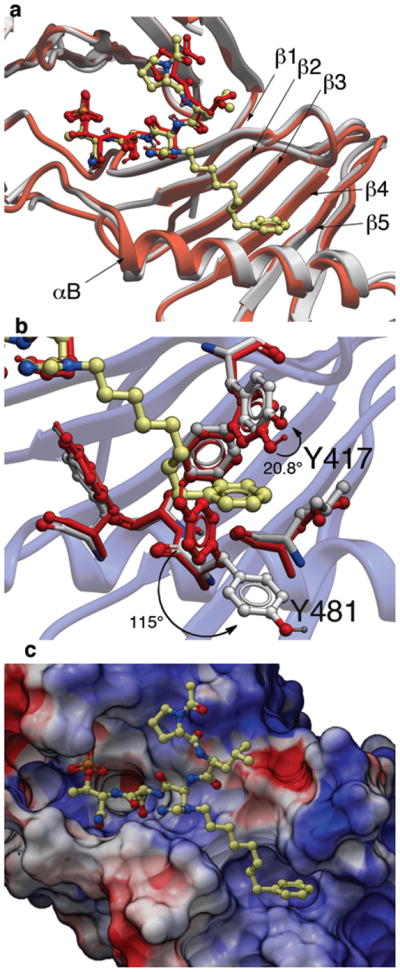
X-ray co-crystal structures of Plk1 PBD complexed with peptides 1 and 4j. (a) PBD in complex with 1 (PBD 3HIK; protein backbone and peptide shown in red) superimposed on the complex with 4j (protein backbone in grey with peptide 4j colored by atom). Key protein structural features are labeled as indicated in reference 21. (b) Plk1 PBD complex with 4j (protein backbone in blue ribbon) showing residue side chains involved with the binding of the C6H5(CH2)8– group of 4j (ligand in yellow with protein carbons in grey) compared with the same residues in the 3HIK structure of PBD-bound 1 (shown in red). Displacements (in degrees) are shown for the Y417 and Y481 phenyl groups. (c) Electrostatic surface of PBD in complex with 4j with coloring based on an arbitrary electrostatic potential scale (positive = blue; negative = red). Peptide 4j is rendered as thick sticks and colored by atom (blue = nitrogen; yellow = carbon; tan = phosphorus and red = oxyben). Graphics were generated using ICM Chemist Pro by Molsoft, Inc. (www.molsoft.com).