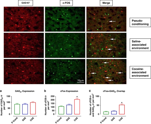
Figure 3.
GAD67 and cFos colocalization after exposure to the cocaine- or saline-associated environment. Photomicrographs of individually immunolabeled GAD67-IR, cFos-IR neurons and double-labeled (GAD67-IR and cFos-IR) neurons (white arrows) in representative animals. Scale bar, 70 μm. (a) Quantification of GAD67 expression/mm2 in pseudo-conditioned animals (control, n=10) and those exposed to the saline- or cocaine-associated environment (SAE, n=10 and CAE, n=10, respectively). (b) Quantification of cFos expression/mm2 in the same groups of animals as in (a). (c) Quantification of cFos–GAD67 overlap/mm2 in the same groups of animals as in (a). Data are expressed as the means±SEM; n=number of animals/experimental group. A greater number of cFos-IR neurons as well as cFos–GAD67 double-labeled neurons were found in animals exposed to cocaine-associated environment. *Indicates significant difference between groups via one-way ANOVA (p⩽0.05).

