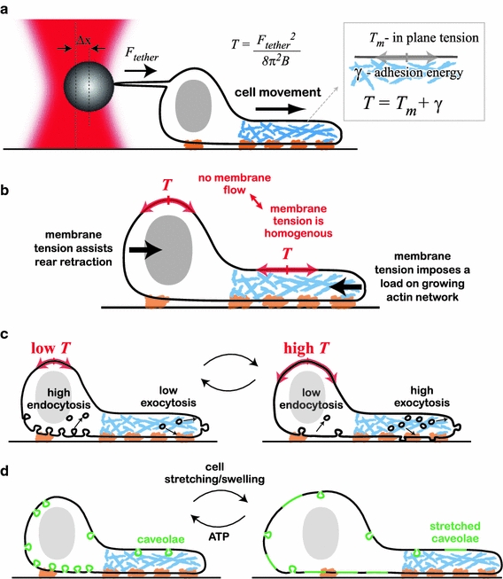
Fig. 3.

Membrane tension in motile cells. a Membrane tension measurements using the tether-pulling assay. A membrane tether is generated by pulling a coated bead away from the cell surface by use of optical tweezers. The force the tether exerts on the bead can be measured from the displacement of the bead from the center of the trap. The apparent membrane tension, which is equal to the sum of the in-plane tension and the adhesion energy per unit area, can be calculated from the measured tether force. b The apparent membrane tension exerts forces perpendicular to the cell boundary (black arrows). In the absence of membrane flow, the apparent membrane tension has to be spatially homogenous. The membrane tension opposes actin network growth at the leading edge, while assisting retraction at the rear. c Endocytosis rates increase with decreasing tension whereas exocytosis rates decrease. It has been suggested that tension-dependent endocytosis and exocytosis are involved in surface area regulation and buffering of membrane tension: increased tension leads to excess exocytosis, leading to an increase in cell surface area and a decrease in tension, and vice versa. d Membrane invaginations, for example caveolae, provide means for rapidly increasing cell surface area and buffering membrane tension. Caveolae require ATP and actin for their synthesis. A rapid increase in tension (because of stretching or swelling of the cells) is buffered by rapid flattening of the caveolae, providing additional surface area
