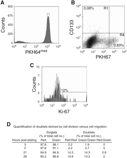
Fig. 7.
Tracking CD133+ cells in monolayer culture. (A) Nonselected primary epithelial cells were stained with PKH67, and a homogeneous subset of PI− PKH67+ cells was isolated using a narrow band of fluorescence. (B) and (C) Cells isolated above were plated onto collagen 1 dishes with stem cell media (SCM) and irradiated STO feeders. After 4 days, the PKH67 cells were stained with anti-CD133-APC (B) and anti-Ki-67 (C). C is the R1 region (CD133+/PKH67−) from B. Cells cultured in the presence of Colcemid® were used to establish the range of fluorescence exhibited by cells that had not divided. Experiments were carried out on 3 patients’ samples. One representative experiment is shown. (D) PKH26 (red)-labeled and PKH67 (green) CD133+ cells were mixed together in a 1:1 ratio and then plated, as above. At 3–26 h, the cells were mapped and red and green cells were counted. To calculate the number of doublets arising from cells division compared to cell migration/aggregation, we counted the number of 2 red cells, 2 green cells, and the number of 1 red and 1 green doublet.

