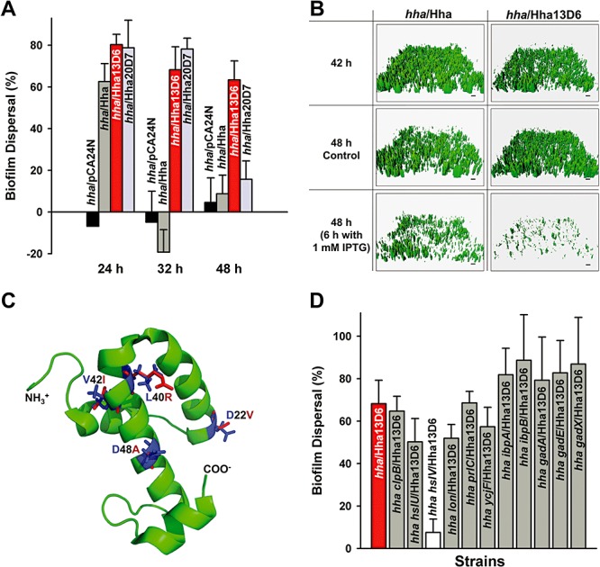
Figure 1.

Biofilm dispersal with Hha13D6. Biofilm dispersal in 96‐well plates for BW25113 hha producing Hha13D6 and Hha20D7 in Luria‐Bertani (LB) glucose (0.2%) at 37°C (A). Biofilm dispersal was quantified by subtracting the normalized biofilm with Hha and its variants produced at 24, 32 and 48 h (8 h after adding 1 mM IPTG) from the normalized biofilm without Hha and its variants produced at 24, 32 and 48 h (no IPTG addition). Each data point is the average of at least two independent cultures, and one standard deviation is shown. Biofilm dispersal in flow‐cells for BW25113 hha producing Hha13D6 from pCA24N (B). Biofilms were formed on glass surfaces in flow‐cells for 42 h then 1 mM IPTG was added for 6 h to induce dispersal (control is no IPTG addition). Scale bar represents 10 µm. Modelled protein structure of Hha13D6 (C). Substituted residues of Hha13D6 (D22V, L40R, V42I and D48A) are shown in red, while the original residues were shown in blue. Impact of ClpB, HslU, HslV, Lon, PrlC, YcjF, IbpA, IbpB, GadA, GadE and GadX on Hha13D6‐mediated biofilm dispersal (D). Biofilm dispersal for cells producing Hha13D6 in LB glucose at 37°C after 32 h (8 h with 1 mM IPTG) in the following hosts: BW25113 hha (hha), BW25113 hha clpB (hha clpB), BW25113 hha hslU (hha hslU), BW25113 hha hslV (hha hslV), BW25113 hha lon (hha lon), BW25113 hha prlC (hha prlC), BW25113 hha ycjF (hha ycjF), BW25113 hha ibpA (hha ibpA), BW25113 hha ibpB (hha ibpB), BW25113 hha gadA (hha gadA), BW25113 hha gadE (hha gadE) and BW25113 hha gadX (hha gadX). Each data point is the average of at least two independent cultures, and one standard deviation is shown.
