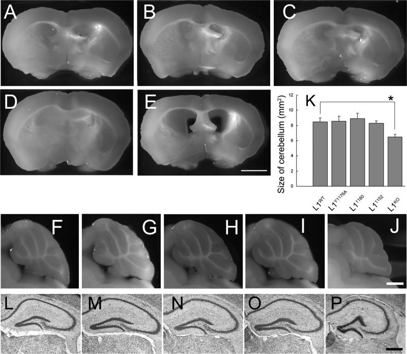
Figure 4. Lack of gross morphological abnormalities in L1CD mutant mice. A, F, L: WT; B, G, M: L1Y1176A; C, H, N: L11180; D. I, O: L11152; E, J, P: L1KO.
(A-E) No hydrocephalus in L1CD mutant mice. Coronal section from brains showed that there are no enlarged ventricles in L1CD mice, although L1KO mice have enlarged ventricles. n = 3. Scale bar, 200 μm
(F-J) Normal morphology of cerebellar lobe in mid-sagittal sections of brains. The cerebellar lobule shows normal morphology in L1CD mutant mice. n=3. Scale bar, 1 mm.
(K) Morphometric analysis of cerebellum at the midline of brains. There is no significant difference in the size of the cerebellum of L1CD mutant mice, although the L1KO mice cerebella are significantly smaller. n = 3. *p < 0.05.
(L-P) Cresyl violet staining for hippocampus region. Formation of hippocampus is normal in L1CD mutant mice compared with L1WT mice, although L1KO mice have a smaller hippocampus. Scale bar = 500 μm

