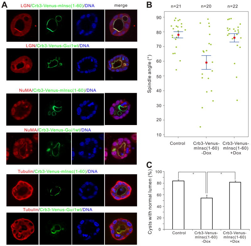
Figure 6. Apical targeting of mInsc(1-60) leads to apical recruitment of endogenous LGN and subsequent spindle misorientation and defective cystogenesis in MDCK cysts.
(A) MDCK cells stably expressing Crb3-Venus-mInsc(1-60) or Crb3-Venus-Gαi1 were cultured in matrigel. After culturing for 4 days in the absence of doxycycline, cysts were fixed and stained with anti-LGN, anti-NuMA or anti-Tubulin antibodies, respectively. DNA was stained with Hoechst 33342. Single confocal images from the middle of the cysts are shown. (B) Scatter diagram of the metaphase spindle angles in control or Crb3-Venus-mInsc(1-60) cysts in the absence (-Dox) or presence (+Dox) of 20 ng/ml of doxycycline. Results were from three independent experiments. Pink dots indicate mean values, green dots indicate individual data points, and error bars represent the SEM of the total number of spindles analyzed. (C) Quantification of cysts with single normal lumen from MDCK cells expressing Venus (control) or Crb3-Venus-mInsc(1-60) in the absence (-Dox) or presence (+Dox) of 20 ng/ml of doxycycline. Values are mean ± SD from three independent experiments, n > 100 cysts/experiment. *P < 0.01. Also see Fig. S6.

