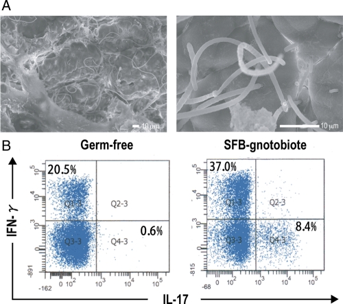
Figure 1.
Morphology and immunostimulatory activity of SFBs grown in SFB-monoassociated gnotobiotic mice. (A) Electron microscopic examination of SFBs. BALB/c germ-free mice were colonized with the SFB, and the terminal ilea collected 14 days after inoculation were examined by scanning electron microscopy. SFBs show a characteristic long filamentous morphology and preferentially colonized around Peyer's patches. Epithelial cell surfaces to which SFB attach are deeply indented. (B) Induction of Th1 and Th17 cells by SFB colonization. Intestinal LPLs were isolated from BALB/c germ-free mice or SFB-monoassociated gnotobiotic mice and the percentages of IFN-γ+ or IL-17+ cells within the CD4+ lymphocyte population in each LPL preparation were determined by FACS analysis.

