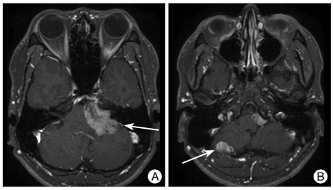
Fig. 1.
A magnetic resonance (MR) image showing two heterogenously enhanced masses in the left cerebellopontine angle (CPA) and the convexity of the right cerebellar hemisphere. The large mass was 4.2 cm in size and well demarcated, compressing the brain stem to the opposite side (A). The small mass was 1.5 cm in size, and the mass effect was not combined (B).

