
Figure 1.
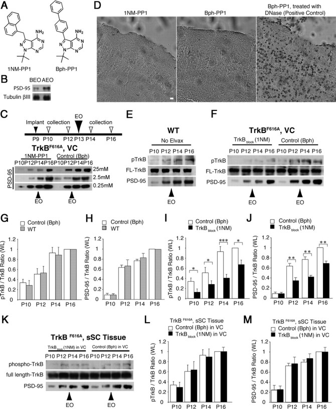
TrkB blockade suppresses developmental increase of PSD-95 synthesis. A, Chemical structure of 1NM-PP1 (left) and Bph-PP1 (right). B, PSD-95 level significantly increased 6 h AEO compared to BEO at P13 in synaptosomal fractions from mouse visual cortex. C, Among three different concentrations, Elvax containing 1NM-PP1 at 25 mm showed the highest suppression of PSD-95 at P12, P14, and P16 compared to Bph-PP1 containing Elvax. The inset diagram (top) in this figure and those in the following figures illustrate the sequence of procedures for the experiments. D, The TUNEL assay showed no apoptosis in VC under the 1NM-PP1-Elvax (left) or Bph-PP1-Elvax (middle). As a positive control, slices were treated with DNase (right). Scale bar, 10 μm. E, During the period bracketing eye opening, in whole lysates of WT mouse VC, PSD-95 and pTrkB increased, whereas full-length TrkB (FL-TrkB) levels remained constant. F, Both pTrkB and PSD-95 levels were suppressed in whole lysates of VC when TrkB was blocked with 1NM-PP1. FL-TrkB levels in the same lanes are used to normalize because their levels remained constant. G, H, Both pTrkB (G) and PSD-95 (H) levels were similar between WT and Bph-PP1-treated control VC. I, J, Both pTrkB (I) and PSD-95 (J) levels were suppressed in whole lysates of VC when TrkB was blocked with 1NM-PP1. K–M, When TrkB was blocked in VC over the interval encompassing the eye opening (P10–P16), both phosphorylated TrkB (K, L) and PSD-95 (K, M) increased normally in the same animals' superficial visual layers of the superior colliculus (sSC), indicating that the blocking effect was restricted to cortex. The blots in C–F and K were first probed for pTrkB, then stripped and probed for PSD-95 and FL-TrkB. *p < 0.05; **p < 0.01; ***p < 0.001. Error bars represent SEM.