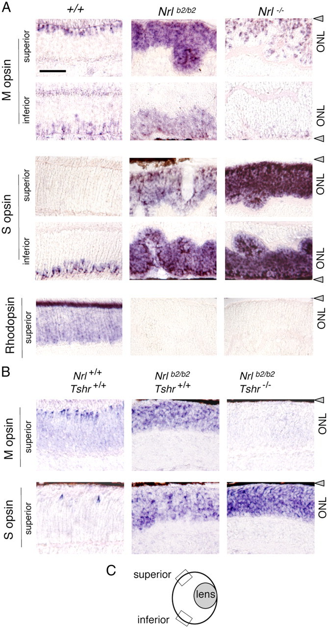
Figure 4.

Differential distribution of M and S opsins in Nrlb2/b2 and Nrl−/− mice. A, In situ hybridization analysis of cone opsin and rhodopsin mRNA in superior and inferior retinal regions in 8-week-old mice. Nrlb2/b2 mice overexpressed M and S opsins with a normal distribution trend, whereas in Nrl−/− mice, S opsin was strongly overexpressed in all retinal regions. Nrl−/− mice showed a small increase in the density of M opsin-positive cells in superior regions compared with +/+ mice, partly due to ONL folding in Nrl−/− mice. B, In situ hybridization analysis of opsin mRNA in hypothyroid Nrlb2/b2 mice at P17. On a hypothyroid Tshr−/− background, M opsin expression was retarded and S opsin overexpression exacerbated in Nrlb2/b2 mice. Scale bar: A (for A, B), 50 μm. C, Diagram of mouse eye section indicating location of superior and inferior retinal fields examined (boxes). Gray triangles, Retinal pigmented epithelium.
