Figure 6.
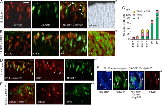
Transient coexpression of TRβ2 and Nrl in photoreceptor precursors. A, Double fluorescence analysis of cells positive for TRβ2 (@TRβ2 antibody, red), NrlpGFP (transgene, green), or both @TRβ2 and NrlpGFP (yellow or orange, arrows in merged image) in the outer neuroblastic layer of +/+ mouse retina at E16.5. Right, Phase contrast image of the same field. Confocal microscope images represent a single 1–1.5 μm z-plane obtained from 10-μm-thick mid-retinal cryosections (same in B–E). For orientation, the gray arrowhead indicates location of retinal pigmented epithelium. B, Double fluorescence analysis of cells positive for TRβ2 and NrlpGFP in +/+ mouse retina during development at E14.5, E18.5, P2, and P8. Doubly positive cells (yellow or orange) were most evident at E16.5–E18.5. Scale bars: A, B, 14 μm. C, Counts of cells positive for TRβ2, GFP, or both TRβ2 and GFP, determined on single z-plane confocal images from experiments shown in A and B. Counts shown were determined in mid-retinal fields. Counts in superior and inferior fields gave similar results. D, Verification of NrlpGFP transgene as a marker for cells expressing endogenous Nrl using an antibody against NRL protein (@Nrl) and direct fluorescence for NrlpGFP in +/+ embryos at E18.5. The @Nrl+ population included almost all GFP+ cells (yellowish and orange cells, merged image on the left) and a few @Nrl+ cells that were negative for GFP (white arrows, middle). E, Independent identification of cells that coexpress TRβ2 (@bGal) and endogenous NRL protein (@Nrl) (yellow or orange cells in merged image, left). Analysis was performed on E18.5 embryos homozygous for a targeted insertion of lacZ in the TRβ2-specific exon of the Thrb gene. Scale bars: D, E, 10 μm F, Immunofluorescence analysis for coexpression of S opsin (@S opsin antibody, blue), NrlpGFP (direct fluorescence, green) and Thrb2p-lacZ transgenes (@bGal antibody, red, indicator for TRβ2) in +/+ mice at P0. Arrows, S opsin/GFP doubly positive cell; arrowheads, S opsin/GFP/bGal triply positive cell; asterisks, S opsin-positive cell with no detectable GFP or bGal. Gray triangle, Retinal pigmented epithelium.

