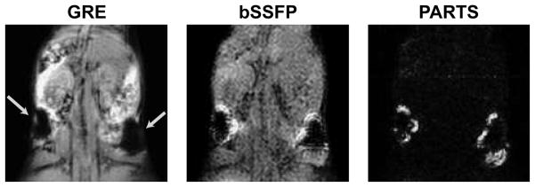
Figure 10.
Coronal projections (over 4 slices) from in vivo GRE, bSSFP, and PARTS acquisitions at 1.5 T. The mouse was injected with 1.5 and 3 million SPIO-labeled cells at two sites (arrows). Separate water- and fat-suppressed data were combined to generate the PARTS image. The suboptimal background suppression with bSSFP compromises the conspicuity of the positive contrast images. On the other hand, the background is almost nonexistent in the PARTS image.

