Figure 9.
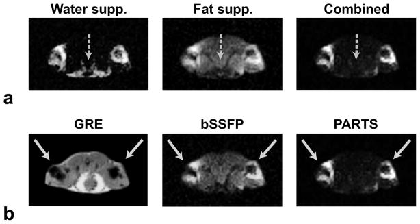
Axial slices from in vivo acquisitions performed at 1.5 T, obtained in a mouse injected with SPIO-labeled cells into its hind limbs. a: The water- and fat-suppressed PARTS acquisitions are separately displayed along with the final combination. The water-suppressed image has residual fat signal (dashed arrow), while the fat-suppressed image has remnant on-resonant signal. Their combination simultaneously suppresses both sources of background signal. b: The GRE, bSSFP and combined PARTS images of the same slice, where the solid arrows pinpoint the injection sites. While bSSFP yields considerable background signal, PARTS produces near-complete background suppression.

