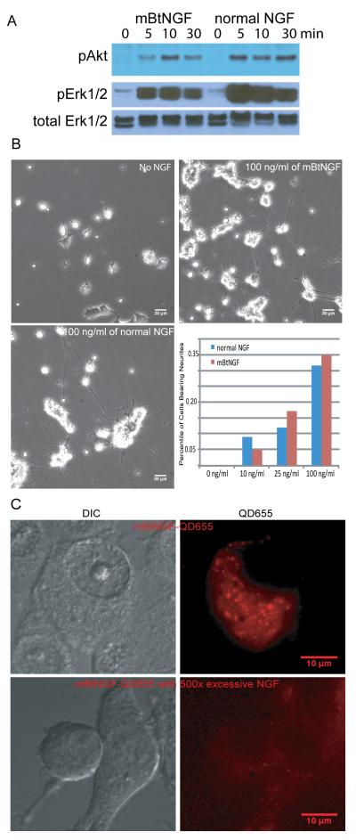
Figure 3.
Analysis of bioactivities of mBtNGF using PC12 cells. A: Activation of Akt and Erk1/2 signaling pathways by mBtNGF. PC12 cells were serum-starved and were stimulated for 0, 5, 10 and 30 min with either 100ng/ml mBtNGF or 100 ng/ml normal NGF. Cell lysates were analyzed by SDS-PAGE/immunoblotting with indicated antibodies. B: Neurite outgrowth assay. PC12 cells were treated with vehicle only (0), or 10, 25 or 100ng/ml mBtNGF or normal NGF at the same concentrations for 6 days. The percentage of cells bearing neurites longer than three times of cell body was presented. Typical micrographs of PC12 cells treated with the vehicle (no NGF), 100 ng/ml mBtNGF or normal NGF were shown. C: Assay for internalization of mBtNGF. PC12 cells after incubating with either 0.2 nM of mBtNGF-QD655 or with 0.2 nM mBtNGF-QD655 together with 100 nM of normal NGF. Both DIC and corresponding fluorescence images were captured and shown.

